What is Vitiligo?
What is Vitiligo?
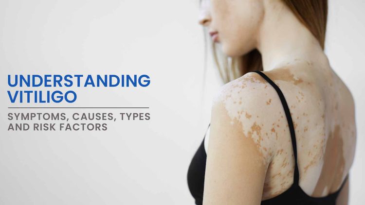
Vitiligo, also called leucoderma or 'white spot' disease, is a condition where there is autoimmune destruction of melanocytes (pigment producing cells of skin), leading to decrease or absence of melanin in the skin, which in turn causes characteristic white patches.
What Other Conditions Look like Vitiligo?
Several other skin conditions can resemble vitiligo in appearance, making accurate diagnosis crucial. Some of these conditions include:
-
Pityriasis alba: Common in children and adolescents, this condition presents as pale, scaly patches on the face, neck, and upper arms. Pityriasis alba patches are typically round or oval and may resemble vitiligo due to their light coloration.
-
Post-inflammatory hypopigmentation: Following skin injuries like burns, cuts, or dermatitis, affected skin may lose pigmentation, resulting in light-coloured patches. This post-inflammatory hypopigmentation can mimic vitiligo but usually occurs in areas of previous trauma or inflammation.
-
Tinea versicolor: Caused by Malassezia yeast, this fungal infection can lead to hypo- or hyperpigmented patches on the skin. Tinea versicolor patches vary in colour from white, pink, tan, or brown and can resemble vitiligo patches.
-
Idiopathic guttate hypomelanosis: This condition presents as small, white, flat spots on sun-exposed skin, often in older individuals. These spots, resulting from decreased melanin production, may be mistaken for vitiligo patches.
-
Halo nevus: A mole (nevus) surrounded by a depigmented ring or halo, caused by an autoimmune reaction against melanocytes in the nevus. This depigmentation around the mole can resemble vitiligo patches.
-
Chemical leukoderma: Exposure to certain chemicals, such as phenols or hydroquinone, can cause depigmented patches on the skin. These patches often occur in areas of skin contact with the offending agent and may mimic vitiligo.
-
Hypopigmented mycosis fungoides: A type of cutaneous T-cell lymphoma that can present with hypopigmented patches on the skin, sometimes resembling vitiligo but often occurring in a more linear or streaky pattern.
-
Piebaldism: A rare genetic disorder characterised by congenital patches of depigmented skin and white hair, typically present from birth. Piebaldism is caused by mutations in the KIT gene, resulting in leucoderma patches that can resemble vitiligo.
What are the Symptoms of Vitiligo?
Vitiligo presents primarily as light-coloured patches or macules on the skin. The most common symptom of vitiligo is the appearance of well-defined, irregularly shaped patches of lighter skin that may be milky-white, white, or lighter in colour than the surrounding skin. Other symptoms and characteristics of vitiligo may include:
-
Progressive enlargement: Vitiligo patches may gradually enlarge and spread over time, affecting larger areas of the skin. The progression of vitiligo can be slow and steady or may occur in episodic bursts of activity followed by periods of stability.
-
Symmetry: In many cases, vitiligo patches have a symmetrical distribution, occurring in corresponding areas on both sides of the body. For example, if a patch develops on one elbow, a similar patch may appear on the other elbow.
-
Well-defined borders: Vitiligo patches typically have well-defined borders, with clear boundaries separating the depigmented skin from the surrounding normal skin. These borders may be irregular or jagged in appearance.
-
Variation in size and shape: Vitiligo patches can vary in size and shape, ranging from small, isolated spots to larger patches that merge together to form larger areas of depigmentation.
-
Involvement of mucous membranes: In some cases, vitiligo may also affect mucous membranes lining the inside of the mouth, nose, genitals, and rectum. Depigmented patches may appear on these mucous membrane surfaces, although this is less common than skin involvement.
It's important to note that vitiligo can vary widely in severity and extent among individuals, with some people experiencing only a few small patches of depigmentation, while others may have widespread involvement affecting large areas of the body.
What are the Types of Vitiligo?
Depending on the extent of involvement it could be of following types:
-
Generalised/Non-segmental vitiligo (NSV): This form of vitiligo is the most common presentation, accounting for about 90% of cases. NSV is characterised by the symmetric distribution of depigmented patches on both sides of the body, which typically develop gradually and may vary in size and shape. Commonly affected areas include the face, hands, arms, feet, and genitalia.
-
Segmental vitiligo: Unlike non-segmental vitiligo, which affects both sides of the body symmetrically, segmental vitiligo is confined to one side of the body or a specific segment. This type often presents at a younger age and tends to stabilise without spreading to other areas.
-
Mixed vitiligo: Mixed vitiligo combines features of both non-segmental and segmental vitiligo. In this type, individuals may exhibit both symmetrical depigmented patches typical of non-segmental vitiligo and unilateral patches characteristic of segmental vitiligo. Mixed vitiligo can present challenges in diagnosis and management due to the combination of different patterns of depigmentation.
-
Focal vitiligo: Focal vitiligo, a subtype of segmental vitiligo, is characterised by isolated depigmented patches that are limited to small areas of the body. These patches may appear randomly and do not necessarily follow a specific distribution pattern or symmetry. Focal vitiligo can affect any part of the body and may involve only a single patch or multiple patches within the same segment.
-
Trichrome vitiligo: Trichrome vitiligo is a rare subtype characterised by the presence of three different colours within depigmented patches. These colours typically include areas of pure white (depigmented skin), areas of normally pigmented skin, and areas of hyperpigmentation (darkened skin). Trichrome vitiligo results from a combination of depigmented, normally pigmented, and hyperpigmented areas within the same vitiligo lesion.
-
Universal vitiligo: Universal vitiligo represents the most severe form of the condition, characterised by extensive and widespread depigmentation affecting most or all of the body's skin surface. In universal vitiligo, depigmented patches may cover large areas of the body, including the face, trunk, extremities, and even the hands, feet, and mucous membranes.
What are the Risk Factors for Vitiligo?
Several factors can increase the risk of developing vitiligo. These include:
-
Genetic predisposition: Family history plays a significant role, as individuals with relatives affected by vitiligo have a higher risk of developing the condition themselves. Genetic predisposition contributes to the autoimmune response that leads to melanocyte destruction in vitiligo.
-
Autoimmune diseases: Vitiligo often coexists with other autoimmune disorders such as Addison’s disease, pernicious anaemia, Grave’s disease, type-1 diabetes, lupus, psoriasis, rheumatoid arthritis, and thyroid disease.
-
Age: While vitiligo can develop at any age, it often first appears before the age of 20, with the majority of cases occurring before the age of 30. However, vitiligo can also develop later in life.
-
Environmental factors: Exposure to certain environmental factors, such as chemicals, trauma to the skin (e.g., sunburn, cuts, burns), and emotional stress, may trigger or exacerbate vitiligo in susceptible individuals. However, the exact triggers of vitiligo remain unclear.
-
Immune system dysfunction: Vitiligo occurs when the immune system mistakenly attacks and destroys melanocytes, the pigment-producing cells in the skin. Dysfunction of the immune system, therefore, contributes to the development of vitiligo.
Understanding these risk factors can help individuals and healthcare providers identify those at higher risk of developing vitiligo and implement preventive measures or early intervention strategies when appropriate.
How is Vitiligo Diagnosed?
Vitiligo is typically diagnosed through a combination of medical history, physical examination, and sometimes additional tests. Here's how the diagnosis is typically made:
-
Medical history: The healthcare provider will begin by taking a detailed medical history, including any symptoms experienced, family history of autoimmune diseases or vitiligo, and any recent changes in skin pigmentation. Information about past medical conditions, medications, and exposure to potential triggers or risk factors may also be relevant.
-
Physical examination: A thorough physical examination of the skin is conducted to assess the presence of depigmented patches characteristic of vitiligo. The doctor examines the skin for well-defined, irregularly shaped patches that may be milky-white, white, or lighter in colour than the surrounding skin. The distribution, size, shape, and symmetry of the patches are also noted.
-
Wood's lamp examination: In some cases, a Wood's lamp examination may be performed to aid in the diagnosis of vitiligo. A Wood's lamp emits ultraviolet (UV) light that can highlight depigmented areas of the skin, making them more visible. In vitiligo, affected areas may appear more distinct under the Wood's lamp compared to normal skin.
-
Laboratory tests: In some cases, additional laboratory tests may be ordered to evaluate thyroid functions, autoimmune markers, or other underlying conditions associated with vitiligo or autoimmune diseases. These tests may include blood tests such as thyroid function tests, antinuclear antibody (ANA) tests, or thyroid antibodies.
-
Skin biopsy: In certain situations, a skin biopsy may be performed to confirm the diagnosis of vitiligo or to rule out other skin conditions with similar features. During a skin biopsy, a small sample of skin tissue is removed and examined under a microscope by a pathologist to assess the presence of melanocytes and any abnormalities.
It's important for individuals with suspected vitiligo to seek evaluation and diagnosis by a qualified dermatologist for appropriate management and treatment.
How is Vitiligo Treated?
While there is no cure for vitiligo, several treatment options are available to help improve skin appearance and reduce the contrast between depigmented and pigmented areas. These treatment options include:
-
Topical corticosteroids: Topical corticosteroids are commonly used as the first-line treatment for localised vitiligo, particularly in areas with active disease or recent onset. These medications help to suppress the immune response and reduce inflammation, promoting repigmentation of the skin. Corticosteroids are typically applied to affected areas once or twice daily for several months.
-
Topical Calcineurin Inhibitors: Calcineurin inhibitors, such as tacrolimus ointment and pimecrolimus cream, are another class of topical medications used to treat vitiligo. These medications work by suppressing the immune response and reducing inflammation in the skin, leading to repigmentation of depigmented areas.
-
Phototherapy (Light Therapy): Phototherapy involves exposing the skin to ultraviolet (UV) light to stimulate melanocyte activity and promote repigmentation. Narrowband ultraviolet B (NB-UVB) phototherapy is the most commonly used form of phototherapy for vitiligo and is often administered two to three times per week by a dermatologist.
-
Depigmentation: In cases of extensive or widespread vitiligo where repigmentation treatments are not feasible or effective, depigmentation therapy may be considered. Depigmentation involves using topical agents, such as monobenzone, to intentionally lighten unaffected areas of the skin to match the depigmented areas.
-
Surgical Treatments: Surgical options may be considered for localised or stable vitiligo that has not responded to other treatments. Surgical treatments for vitiligo include techniques such as autologous melanocyte transplantation, punch grafting, suction blister grafting, and split-thickness skin grafting. These procedures involve transplanting melanocytes or skin from unaffected areas to depigmented areas.


